 |
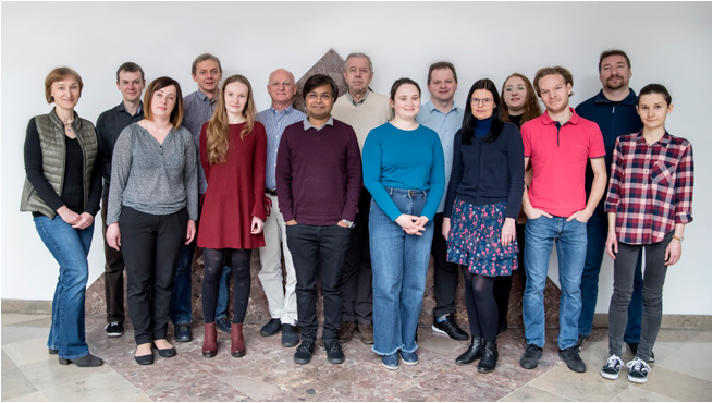
Jacek Kuźnicki, PhD, Professor
Correspondence address:
Laboratory of Neurodegeneration
International Institute of Molecular and Cell Biology
4 Ks. Trojdena Street, 02-109 Warsaw, Poland
Email: This email address is being protected from spambots. You need JavaScript enabled to view it.
|
DEGREES
1993 - Professor, awarded by the President of the Republic of Poland
1987 - DSc. Habil., Nencki Institute of Experimental Biology PAS, Warsaw, Poland
1980 - PhD in biochemistry, Nencki Institute of Experimental Biology PAS, Warsaw, Poland
1976 - MSc in biochemistry, Faculty of Biology, University of Warsaw, Poland
PROFESSIONAL EXPERIENCE
2001-present - Professor, Head of Laboratory of Neurodegeneration, International Institute of Molecular and Cell Biology in Warsaw, Poland
2001-2018 - Director, International Institute of Molecular and Cell Biology in Warsaw, Poland; Feb-Dec 2018 Acting Director, International Institute of Molecular and Cell Biology in Warsaw, Poland
2017-present - Deputy Chair of the Council of Provosts, 2nd Division: Biological and Agricultural Sciences, Polish Academy of Sciences
2000-2001 - Director, Centre of Excellence Phare Sci-Tech II, Nencki Institute of Experimental Biology, Polish Academy of Sciences, Warsaw, Poland
1999-2001 - Acting Director, International Institute of Molecular and Cell Biology in Warsaw, Poland; Organizer and Director, Centenarian Program
1996-2002 - Head, Laboratory of Calcium Binding Proteins, professor 2002- 2014 Nencki Institute of Experimental Biology, Polish Academy of Sciences, Warsaw, Poland
1992-1995 - Visiting Professor, NIMH, Bethesda, MD, USA
1991-1992 - Deputy Scientific Director, Nencki Institute of Experimental Biology, Polish Academy of Sciences, Warsaw, Poland
1986-1992 - Associate Professor and Head of Laboratory of Calcium Binding Proteins, Nencki Institute of Experimental Biology, Polish Academy of Sciences, Warsaw, Poland
1984-1985 - Research Associate, Nencki Institute of Experimental Biology, Polish Academy of Sciences, Warsaw, Poland
1981-1984 - Visiting Fellow, NIH, Bethesda, MD, USA
1980-1981 - Post-doctoral Fellow, Nencki Institute
1976-1980 - PhD Student, Nencki Institute of Experimental Biology PAN, Warsaw
PROFESSIONAL TRAINING
2018 - Visiting Professor, Laboratory of H. Burgess, National Institute of Child Health and Human Development, Bethesda, MD, USA
2015 - Visiting Professor, Laboratory of W. Harris, University of Cambridge, UK
2014 - Visiting Professor, Laboratory of B.E. Snaar-Jagalska, Leiden University, The Netherlands
1992-1995 - Visiting Professor, Laboratory of D. Jacobowitz, National Institute of Mental Health, Bethesda, MD, USA
1981-1984 - Visiting Fellow (postdoc), Laboratory of E.D. Korn, National Institute of Heart, Lung and Blood, Bethesda, MD, USA
MEMBERSHIP IN SCIENTIFIC SOCIETIES, ORGANIZATIONS, AND PANELS
2020 - Ordinary Member, Polish Academy of Sciences
2018-2022 - Member, Council of the National Science Centre and Chair of International Commission
2017-2018 - Deputy Chair, Council of Provosts, 2nd Division, Polish Academy of Sciences
2016-present - Member, International Advisory Board, Małopolska Centre of Biotechnology, Jagiellonian University
2011-present - Member, International Expert Council of the Research and Education Center, State Key Laboratory of Molecular and Cellular Biology, Ukraine
2011-2014 - Member, Science Policy Committee, and Rotating President (Jul-Dec 2012), Ministry of Science and Higher Education
2008-present - Board Member, European Calcium Society
2008-2018 - Member, Board of Directors, and Rotating President (Jul-Dec 2016, Jul-Dec 2013, Jul-Dec 2010), Biocentrum-Ochota Consortium
2006-2011 - Member, Advisory Group, 7FP HEALTH, European Commission
2004-2019 - Corresponding Member, Polish Academy of Sciences
2004-present - Honorary Chair and co-founder, BioEducation Foundation
2002-present - Head of Program Board, Centre for Innovative Bioscience Education
1993-2014 - Member, Scientific Council, Nencki Institute of Experimental Biology, Polish Academy of Sciences
1996-1998 & 2000-2002 - Vice-President, Biotechnology Committee, Polish Academy of Sciences
1989 - 1991 - General Secretary, Polish Biochemical Society
HONORS, PRIZES AND AWARDS
2018 - Commemorative medal for the 100th Anniversary of the Nencki Institute for outstanding contribution to the development of this Institute, supporting its activities, shaping the image and building a mutual success
2013 - Crystal Brussels Prize for outstanding achievements in 7th Framework Programme of the European Union for Research and Development
2013 - Award from the 2nd Division of Biological and Agricultural Sciences, Polish Academy of Sciences for series of works on β-catenin
2011 - Konorski Award for the best Polish research work in neurobiology awarded by the Polish Neuroscience Society and Committee on Neurobiology of the PAS, Warsaw
2008 - Officer's Cross of the Order of Polonia Restituta awarded by the President of Poland
2004-2007 - Professorial Subsidy Program Award from Foundation for Polish Science
2003 - Prime Minister Award for the scientific achievements, Warsaw
2001 - Award from the Division of Biological Sciences of the PAS for the work on CaBP
1998 - Knight's Cross of the Order of Polonia Restituta awarded by the President of Poland
1987 - Polish Anatomical Society Award for the article in “Advances in Cell Biology”
1986 - Skarzynski Award from the Polish Biochemical Society for the best review article in “Advances in Biochemistry”, Warsaw
1977 - Mozolowski Award, Polish Biochem. Society for outstanding Polish young biochemist
1977 - Parnas Award from Polish Biochem. Society for the best paper in biochemical research
1976 - MSc, Magna cum laude, University of Warsaw
DOCTORATES DEFENDED UNDER LAB LEADER’S SUPERVISION
A. Filipek, J. Kordowska, U. Wojda, J. Hetman, M. Palczewska, M. Nowotny, K. Billing-Marczak, Ł. Bojarski, W. Michowski, K. Misztal, M. Figiel, K. Honarnejad, A. Jaworska, K. Gazda.
DESCRIPTION OF CURRENT RESEARCH
We are interested in the molecular mechanisms that are involved in neurodegeneration, with a special emphasis on the role of Ca2+ homeostasis and signaling. These processes are being studied at the genomic, proteomic, and cellular levels using mostly zebrafish, rats, and mice as model organisms. The projects are focused on proteins that are involved in store-operated Ca2+ entry (SOCE) and Ca2+ homeostasis in mitochondria, the involvement of potassium channels in the brain ventricular system, and the in vivo analysis of Ca2+ homeostasis in neurons using zebrafish models. For recent reviews, see Wegierski and Kuznicki (Cell Calcium, 2018) and Winata and Korzh (FEBS Lett, 2018).
ROLE OF STIM PROTEINS IN STORE-OPERATED Ca2+ ENTRY IN NEURONS
We previously showed that STIM1 is involved in a thapsigargin-induced SOCE-like process, whereas STIM2 is mostly active after the EGTA-driven depletion of extracellular Ca2+ (Gruszczynska-Biegala et al., PLoS One, 2011; Gruszczynska-Biegala and Kuznicki, J Neurochem, 2013). We searched for new partners of STIMs other than ORAI channels and found that endogenous STIMs associate with GluA subunits of AMPA receptors (Gruszczynska-Biegala et al., Front Cell Neurosci, 2016). STIM proteins also associate in vitro with NMDA receptors. The results suggest cross-talk between STIM proteins and NMDA receptors and their effect on Ca2+ influx through NMDA receptors (Gruszczynska-Biegala et al., Cells, 2020). Using zebrafish as a model we study STIM2 functions in vivo. We evaluated the expression of Calcium Toolkit genes in zebrafish brain and established the level of SOCE-components (Wasilewska et al., Genes, 2019). We generated stim2a, stim2b, and stim2a:2b knock-out zebrafish lines and analyze them using in vivo calcium imaging in cytosols and mitochondria, behavioral tests and scRNA-Seq.
DYSREGULATION OF Ca2+ HOMEOSTASIS IN COMMON NEURODEGENERATIVE DISEASES
We have been testing the hypothesis that brain dysfunction during aging is induced by changes in Ca2+ homeostasis, which may predispose the brain to SAD pathologies. Transgenic mice that overexpressed key SOCE proteins (STIM1, STIM2, and ORAI1) specifically in brain neurons under the Thy1 promoter were generated. Characterization of the STIM1 line (Majewski et al., BBA Mol Cell Res, 2017, Gruszczynska-Biegała et. al., Cells, 2020), STIM2/ORAI1 (Majewski et al., IJMS, 2020) and ORAI1 lines (Maciag et al., BBA Mol Cell Res, 2019; Majewski et. al., IJMS, 2019) has been reported. Strikingly, aged transgenic ORAI1 mice developed spontaneous seizure-like events that could be observed only in females, suggesting a novel, sex-dependent role of ORAI1 in neural function (Maciag, et al., BBA-MCR 2019). Furthermore, based on RNAseq gene expression profiling analysis and ddPCR conducted on hippocampus we identified a downregulation of Arx gene. The loss of function mutations of ARX are known to be implicated in human cases of epilepsy (Majewski et al., IJMS, 2019).
FAD mutations in presenilins were shown to alter both endoplasmic reticulum (ER) calcium signaling and SOCE, but the role of amyloid precursor protein (APP) and APP FAD mutants in intracellular calcium homeostasis is controversial. Our data indicate that APP and APP FAD mutants are not directly involved in SOCE (Wegierski et al., Biochem Biophys Res Commun, 2016). Instead, we found that APP knockdown resulted in an elevation of the resting levels of ER Ca2+, reduced Ca2+ leakage, and delayed the translocation of STIM1 to Orai1 upon ER Ca2+ store depletion. Our data suggest a regulatory role for APP in ER Ca2+ (Gazda et al., Sci Rep, 2017).
Using quantitative PCR, we compared microRNA (miRNA) profiles in blood plasma from mild cognitive impairment-AD patients (whose diagnoses were confirmed by cerebrospinal fluid [CSF] biomarkers) with AD patients and non-demented, age-matched controls. We adhered to standardized blood and CSF assays that are recommended by the JPND BIOMARKAPD consortium. Six miRNAs (three not yet reported in the context of AD and three reported in AD blood) were selected as the most promising biomarker candidates that can differentiate early AD from controls with the highest fold changes (Nagaraj et al., Oncotarget, 2017; patent pending: PCT/IB2016/052440).
A loss-of-function mutation of PINK1 causes early-onset Parkinson’s disease in humans. In collaboration with Oliver Bandmann (University of Sheffield), we used a pink1 mutant (pink1-/-) zebrafish line to study alterations of Ca2+ homeostasis (Flinn et al., Ann Neurol, 2013; Soman et al., Eur J Neurosci, 2016). We generated mcu knock-out fish, which is viable and fertile. The pink1-/-/mcu-/- double-knockout line shows no loss of dopaminergic neurons, suggesting that Ca2+ that enters mitochondria via the mitochondrial Ca2+ uniporter is involved in the pathology of the pink1 mutant. We expressed a mitochondrial Ca2+ probe (CEPIA2mt) under a pan-neuronal promoter (elavl3) to visualize Ca2+ levels in the mitochondrial matrix of a zebrafish (Tg[elavl3:CEPIA2mt]). Lightsheet fluorescence microscopy enabled us to visualize chemically inducible Ca2+ flux in zebrafish neurons in vivo. Inactivation of mitochondrial calcium uniporter (mcu) is protective in both genetic and chemical models of Parkinson disease (Soman et al., Biol Open. 2019). Hence, regulating the mcu function could be an effective therapeutic target in managing pathological changes in the Parkinson disease.
RARE AND ULTRA-RARE DISEASES WITH NEUROLOGICAL PRESENTATION
We previously showed that a mutation of huntingtin (HTT) in YAC128 mice (i.e., a model of Huntington’s disease) resulted in the higher expression of some components of the calcium signalosome, including huntingtin-associated protein 1 (HAP1) and calcyclin (S100A6) binding protein (CacyBP/SIP; Czeredys et al., Front Mol Neurosci, 2013). We detected greater activity of SOC channels in medium spiny neurons (MSNs) from YAC128 mice and found that some tetrahydrocarbazoles restored the disturbances in Ca2+ homeostasis and stabilized SOCE in YAC128 MSN cultures (Czeredys et al., Biochem Biophys Res Commun, 2017). The overexpression of HAP1 protein isoform A in MSNs from YAC128 mice was found to enhance the activity of SOC channels after the activation of IP3R1 (Czeredys at al., Front Cell Neurosci, 2018).
We also study Ca2+ homeostasis and its impact on the progression of neurodegeneration, behavioral changes, seizures, and sleep disturbances in several rare neurodegenerative diseases affecting children. Using CRISPR-Cas9 technology, we have created zebrafish lines with a mutation in the npc2, sgsh, ppp3ca, ptpn4a, and ptpn4b genes. We use those lines for identification of new diagnostic markers for early diagnosis and monitoring progression of two lysosomal storage diseases - the Niemann-Pick type C (NPC), and mucopolisaccharidosis type III (MPS-III), as well better understanding mechanisms leading to pathological changes in two ultra-rare conditions affecting just a few children world-wide (Rydzanicz et al., Eur J Hum Genet., 2018; Szczałuba et al., Clin Genet, 2019). The Zebrafish model is especially suited for drug screens. As NPC and MPS-III share many futures with Alzheimer's disease, hence drug targets that will be identified might find application in the management of a broad range of common neurodegenerative diseases.
DEVELOPMENT OF HOLLOW ORGANS
Subunits of the voltage-gated potassium channels Kcnb1 (Kv2.1) and Kcng4 (Kv6.4) are expressed in several hollow organs (e.g., brain ventricular system [BVS], ear, and eye) where they form tetrameric K+ channels and antagonize each other’s activity. The deficiency of Kcnb1 in zebrafish causes microcephaly, and the gain-of-function of Kcnb1 causes hydrocephalus. Kcng4 acts in a reverse manner (Shen et al., Sci Rep, 2016). The deficiency of BVS in human causes epilepsy (Jedrychowska and Korzh, Dev Dynam, 2019). Formation of the BVS occurs during the early neural development of vertebrates (Korzh, Cell Mol Life Sci, 2018). Deficiency of the BVS has been linked to numerous neurodegenerative diseases. Formation of the BVS depends on many factors, including the ependyma (i.e., cells that line the BVS cavity) and circumventricular organs, including the choroid plexus (Garcia-Lecea et al., Front Neuroanat, 2017; Korzh and Kondrychyn, Semin Cell Dev Biol, 2019). To study the role of K+ channels in the development of hollow organs, we generated a mutant of Kcnb1 and two mutants of kcng4b with deficiency in the BVS and ear. To distinguish the mechanism of action of KCNB1 mutations in human we initiated an analysis of developmental defects caused by mRNA of these mutants in the developing brain and ear of zebrafish


