Notice: Trying to get property of non-object in /home/users/iimcb/public_html/test/bip/modules/mod_menu/mod_menu.php on line 20


  • Laboratory of Structural Biology

Laboratory of Structural Biology

 bochtler m

Matthias Bochtler, PhD, Professor 

Correspondence address:
Laboratory of Structural Biology
International Institute of Molecular and Cell Biology
4 Ks. Trojdena Street, 02-109 Warsaw, Poland
Email: This email address is being protected from spambots. You need JavaScript enabled to view it.
Office: This email address is being protected from spambots. You need JavaScript enabled to view it.
tel: +48 (22) 597 0732; fax: +48 (22) 597 0715

DEGREES

2009 - Professor of Biological Sciences, nomination by the President of the Republic of Poland

2006 - DSc Habil, Institute of Bioorganic Chemistry, Polish Academy of Sciences, Poznań, Poland

1999 - PhD in Biochemistry, Technical University of Munich, Germany

1995 - MSc in Experimental Physics, Munich University, Germany

PROFESSIONAL EMPLOYMENT

2011-present - Professor, Head of Laboratory of Structural in Biology, International Institute of Molecular and Cell Biology in Warsaw, Poland, and Laboratory of Genome Engineering, Institute of Biochemistry and Biophysics, Polish Academy of Sciences,Warsaw, Poland

2007-2011 - Part-time Director of Structural Biology, Cardiff University, United Kingdom

2001-2010 - Head, Joint MPG-PAS Junior Research Group, International Institute of Molecular and Cell Biology in Warsaw, Poland

2000 - Patent training, Weickmann & Weickmann

1999-2000 - Postdoctoral Fellow, Max Planck Institute of Biochemistry, Martinsried, Germany

RESEARCH TRAINING

1996-1999 - Research Assistant, Max Planck Institute of Biochemistry, Martinsried, Germany

1995-1996 - Internship, Medical Microbiology, University of Regensburg,  Germany

1992-1993 - Guest Student, Cambridge University, United Kingdom

1990-1992 - Studies in Physics, Munich University, Germany

HONORS, PRIZES AND AWARDS

2018 - TEAM Foundation for Polish Science

2018 - International Academic Partnerships Programme, Polish National Agency for Academic Exchange

2018 - DAINA, National Science Centre

2015 - HARMONIA, National Science Centre

2012 - MAESTRO, National Science Centre

2011 - TEAM, Foundation for Polish Science Professor Stefan Pieńkowski Award

2004 - EMBO/HHMI Young Investigator Award

2000 - Crystal Award, Germany

1998 - Crystal Award Germany

1990 – 1992 - Scholarship from Deutsche Studienstiftung and Bavarian State

DOCTORATES DEFENDED UNDER LAB LEADER’S SUPERVISION

R. Filipek, M. Firczuk, M. Lipka, R. Szczepanowski, M. Kaus-Drobek, M. Sokołowska, G. Chojnowski, H. Korza, M. Wojciechowski, W. Siwek, P. Haniewicz, A.A. Kazrani, K. Mierzejewska.

The prevalent DNA modification in eukaryotes is C5-cytosine methylation. Biophysically, this modification stabilizes double-stranded DNA. Evolution has built on and enhanced this tendency to utilize C5-methylation, which predominantly occurs in the symmetric CpG context for transcription control. Consistent with its biophysical effects, methylation in transcription control structures (e.g., promoters) represses transcription; methylation elsewhere (e.g., gene bodies) enhances transcription by suppressing aberrant initiation. Methylation can be introduced in one step by de novo and maintenance methyltransferases and propagated by feed-forward loops that link DNA methylation and repressive chromatin marks. Methylation is most easily lost passively as a result of DNA replication, but it can also be actively erased through a mechanism that utilizes TET catalyzed oxidation to prime DNA for base excision repair. The genetics and cell biology of DNA methylation are unique to eukaryotes, but the biochemistry of DNA methylation (and to some extent also DNA demethylation) is also conserved in prokaryotes. We seek to answer biochemical questions using more robust bacterial proteins and answer genetic/ cell biological questions using zebrafish models and human genetic data (e.g., for malignancies with defects in demethylation machinery).

METHYLATION SENSING 

In 2019, we continued our research on relatively well-behaved prokaryotic model proteins to study the specific recognition of 5-methylcytosine and its oxidized congeners in DNA. The repertoire of 5mC and 5hmC binding proteins is relatively small, and most proteins that specifically bind DNA with these bases contain the same domains. Domains that bind fully methylated DNA in the context of CpG include zinc finger and MBD domains. In contrast, hemi-methylated DNA is typically bound by SRA domains. During the last year, we considerably broadened the repertoire of known domains that recognize 5mC and 5hmC. SRA domains belong to the larger superfamily of PUA domains, which also comprises PUA domains in the strict sense, ASCH domains, EVE domains, and several other lesser known domain groups. To the extent that function was known, a clear division of labor appeared to be in place. SRA domains were associated with the binding of modified DNA, whereas other families within the PUA superfamily either were known to be involved in the binding or processing of modified RNA or had completely unknown functions. In silico screens that were performed in collaboration with Dr. Shuang-Yong Xu (New England Biolabs) showed that many PUA superfamily domains in bacteria are fused to endonucleolytic domains that are associated with DNA cleavage. Subsequent biochemical experiments demonstrated that the fusion proteins indeed cleaved modified DNA, although not with the same degree of specificity as SRA domains (Lutz et al., Nucleic Acids Res, 2019). We also crystallographically characterized a few prototype enzymes and solved their structures with and without DNA. The structures illustrate the mode of recognition of methyl- or hydroxymethyl modifications. Similar DNA modification-sensing domains exist in eukaryotes and have been implicated in malignancies, but their biochemical behavior requires further investigation. NEco is the modification-sensing domain of EcoKMcrA, which is one of the earliest studied restriction endonucleases of E. coli. EcoKMcrA efficiently restricts DNA that contains 5mC or 5hmC, provided the modifications are present in the right context. Efficiency is much greater for fully methylated DNA than for hemi-methylated DNA. Our previous work indicated that the NEco modification-sensing domain was phylogenetically unrelated to other methylationsensing domains. In 2019, we elucidated the binding mode of the domain to modified DNA and found that modification sensing is also locally very different from previous observations (Slyvka et al., Nucleic Acids Res, 2019). To date, NEco has been shown to be very good at discriminating (hydroxy)methylated from unmethylated DNA. Further research will likely expand the currently known and narrowly defined phylogenetic distribution (Fig. 1).

fig.1

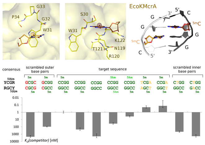
fig.1. Modification-dependent DNA binding by the N-terminal domain of EcoKMcrA endonuclease (NEco). (Top) Interaction between fully modified DNA with (hydroxy)methyl binding pockets of EcoKMcrA. (Bottom) Sequence and modification specificity of EcoKMcrA N-terminal domain determined by EMSA competition experiments (for experimental details, see Slyvka et al., Nucleic Acids Res, 2019).

DEMETHYLATION 

In contrast to methylation sensing, demethylation has no clear equivalent in prokaryotes and thus needs to be studied using eukaryotic models. Our research primarily focuses on the ways in which TET proteins identify their targets. Two of our collaborators, Dr. Tomasz Jurkowski (Cardiff University, United Kingdom) and Dr. Tim Hore (Otago University, New Zealand), provided strong biochemical evidence that the locus specificity of TETs is at least partially attributable to sequence specificity. We solved structures with preferred and discriminated substrates to better understand the mode of sequence recognition. Based on the lack of sequence-specific contacts between TETs and their target DNAs outside the CpG core recognition sequence, we initially hypothesized that DNA bending was responsible for sequence specificity. Our crystal structures do not support this hypothesis, however, and instead reveal an unexpected mechanism of sequence recognition that also explains similarities in preference of different TET paralogues. We are also continuing our work on links between DNA reprogramming and DNA repair. Some of our experiments, such as investigating the role of NEIL1 and TDG in the excision of oxidized 5-methylcytosine bases, are consistent with the general paradigm that DNA reprogramming co-opts DNA repair. However, based on much circumstantial evidence and the work of others, we suspect that the converse may also be true and that DNA repair may use intermediates that are normally associated with reprogramming. We will test this hypothesis both biochemically and bioinformatically.


bochtler lab

Lab Leader:

  • Matthias Bochtler, PhD, Professor

Senior Researcher:

  • Honorata Czapińska, PhD, DSc Habil

Postdoctoral Researchers:

  • Marek Wojciechowski, PhD


PhD Students:

  • Igor Helbrecht, MSc

  • Magdalena Klimczak, MSc

  • Norbert Osiński, MSc

  • Michał Pastor, MSc

  • Abhishek Pateria, MSc

  • Dominik Rafalski, MSc

  • Anton Slyvka, MSc

  • Anna Stroynowska-Czerwińska, MSc

  • Katarzyna Krakowska, MSc


Another co-worker:

  • Anna Fedenko, MSc


Lab Technician:

  • Agnieszka Olszewska (part-time)


Laboratory Support Specialist:

  • Aleksandra Jakielaszek, MSc Eng.


Contact:

This email address is being protected from spambots. You need JavaScript enabled to view it.

2021

Czapinska H, Winiewska-Szajewska M, Szymaniec-Rutkowska A, Piasecka A, Bochtler M, Poznański J.

Halogen Atoms in the Protein-Ligand System. Structural and Thermodynamic Studies of the Binding of Bromobenzotriazoles by the Catalytic Subunit of Human Protein Kinase CK2.

Pastor M, Czapinska H, Helbrecht I, Krakowska K, Lutz T, Xu S, Bochtler M.

Crystal structures of the EVE-HNH endonuclease VcaM4I in the presence and absence of DNA.

Bochtler M, Fernandes H.

DNA adenine methylation in eukaryotes: Enzymatic mark or a form of DNA damage?

2020

Xu G-L, Bochtler M.

Reversal of nucleobase methylation by dioxygenases.

Fricke T, Smalakyte D, Lapinski M, Pateria A, Weige C, Pastor M, Kolano A, Winata C, Siksnys V, Tamulaitis G, Bochtler M.

Targeted RNA Knockdown by a Type III CRISPR-Cas Complex in Zebrafish.

Lutz T, Czapinska H, Fomenkov A, Potapov V, Heiter DF, Cao B, Dedon P, Bochtler M, Xu S.

Protein Domain Guided Screen for Sequence Specific and Phosphorothioate-Dependent Restriction Endonucleases.

Kisiala M, Kowalska M, Pastor M, Korza HJ, Czapinska H, Bochtler M.

Restriction endonucleases that cleave RNA/DNAheteroduplexes bind dsDNA in A-like conformation.

Skowronek KJ, Bochtler M.

In Vitro Directed Evolution of a Restriction Endonuclease With More Stringent Specificity.

Bochtler M.

Arrhenius-law-governed homo- and heteroduplex dissociation.

2019

Lutz T, Flodman K, Copelas A, Czapinska H, Mabuchi M, Fomenkov A, He X, Bochtler M, Xu S.

A protein architecture guided screen for modification dependent restriction endonucleases.

Mitkowski P, Jagielska E, Nowak E, Bujnicki JM, Stefaniak F, Niedziałek D, Bochtler M, Sabała I.

Structural bases of peptidoglycan recognition by lysostaphin SH3b domain.

Czapinska H, Siwek W, Szczepanowski RH, Bujnicki JM, Bochtler M, Skowronek KJ.

Crystal Structure and Directed Evolution of Specificity of NlaIV Restriction Endonuclease.

Slyvka A, Zagorskaitė E, Czapinska H, Sasnauskas G, Bochtler M.

Crystal structure of the EcoKMcrA N-terminal domain (NEco): recognition of modified cytosine bases without flipping.

2018

Bennabi I, Quéguiner I, Kolano A, Boudier T, Mailly P, Verlhac MH, Terret ME.

Shifting meiotic to mitotic spindle assembly in oocytes disrupts chromosome alignment.

Bochtler M, Mizgalska D, Veillard F, Nowak ML, Houston J, Veith P, Reynolds EC, Potempa J.

The Bacteroidetes Q-Rule: Pyroglutamate in Signal Peptidase I Substrates.

Tamulaitiene G, Manakova E, Jovaisaite V, Tamulaitis G, Grazulis S, Bochtler M, Siksnys V.

Unique mechanism of target recognition by PfoI restriction endonuclease of the CCGG-family.

Stroynowska-Czerwinska A, Piasecka A, Bochtler M.

Specificity of MLL1 and TET3 CXXC domains towards naturally occurring cytosine modifications.

Fernandes H, Czapinska H, Grudziaz K, Bujnicki JM, Nowacka M.

Crystal structure of human Acinus RNA recognition motif domain.

Kisiala M, Copelas A, Czapinska H, Xu S, Bochtler M.

Crystal structure of the modification-dependent SRA-HNH endonuclease TagI

Czapinska H, Kowalska M, Zagorskaite E, Manakova E, Slyvka A, Xu SY, Siksnys V, Sasnauskas G, Bochtler M.

Activity and structure of EcoKMcrA.

Piasecka A, Czapinska H, Vielberg MT, Szczepanowski RH, Kiefersauer R, Reed S, Groll M, Bochtler M.

The Y. bercovieri Anbu crystal structure sheds light on the evolution of highly (pseudo)symmetric multimers.

2017

Slyvka A, Mierzejewska K, Bochtler M.

Nei-like 1 (NEIL1) excises 5-carboxylcytosine directly and stimulates TDG-mediated 5-formyl and 5-carboxylcytosine excision.

Perycz M, Krwawicz J, Bochtler M.

A TALE-inspired computational screen for proteins that contain approximate tandem repeats.

2016

Bochtler M, Kolano A, Xu G-L

DNA demethylation pathways: Additional players and regulators

Szychowska M, Siwek W, Pawolski D, Kazrani AA, Pyrc K, Bochtler M

Type III CRISPR complexes from Thermus thermophilus

Bochtler M., Piasecka A

Haloferax volcanii UbaA, catalytic engine for sampylation and sulfur transfer

Mierzejewska K, Bochtler M, Czapinska H

On the role of steric clashes in methylation control of restriction endonuclease activity

Haniewicz P, Floris D, Farci D, Kirkpatrick J, Loi MC, Büchel C, Bochtler M, Piano D

Isolation of Plant Photosystem II Complexes by Fractional Solubilization

2015

Grabowska M, Jagielska E, Czapinska H, Bochtler M, Sabala I

High resolution structure of an M23 peptidase with a substrate analogue

Burmistrz M, Dudek B, Staniec D, Rodriguez Martinez JI, Bochtler M, Potempa J, Pyrc K

Functional Analysis of Porphyromonas gingivalis W83 CRISPR-Cas Systems

2014

Mierzejewska K, Siwek W, Czapinska H, Kaus-Drobek M, Radlinska M, Skowronek K, Bujnicki JM, Dadlez M, Bochtler M

Structural basis of the methylation specificity of R.DpnI

Jaremko M, Jaremko L, Nowakowski M, Wojciechowski M, Szczepanowski RH, Panecka R, Zhukov I, Bochtler M, Ejchart A

NMR structural studies of the first catalytic half-domain of ubiquitin activating enzyme

Kazrani AA, Kowalska M, Czapinska H, Bochtler M

Crystal structure of the 5hmC specific endonuclease PvuRts1I

Wojciechowski M, Rafalski D, Kucharski R, Misztal K, Maleszka J, Bochtler M, Maleszka R

Insights into DNA hydroxymethylation in the honeybee from in-depth analyses of TET dioxygenase

Sabala I, Jagielska E, Bardelang PT, Czapinska H, Dahms SO, Sharpe JA, James R, Than ME, Thomas NR, Bochtler M

Crystal structure of the antimicrobial peptidase lysostaphin from Staphylococcus simulans

Gallagher JM, Yamak A, Kirilenko P, Black S, Bochtler M, Lefebvre C, Nemer M, Latinkić BV

Carboxy terminus of GATA4 transcription factor is required for its cardiogenic activity and interaction with CDK4

2013

Haniewicz P, De Sanctis D, Büchel C, Schröder WP, Loi MC, Kieselbach T, Bochtler M, Piano D

Isolation of monomeric photosystem II that retains the subunit PsbS

Wojciechowski M, Czapinska H, Bochtler M

CpG Underrepresentation and the Bacterial CpG Specific DNA Methyltransferase M.MpeI

2012

Bochtler M

Structural basis of the TAL effector-DNA interaction

Sabala I, Jonsson IM, Tarkowski A, Bochtler M

Anti-staphylococcal activities of lysostaphin and LytM catalytic domain

Siwek W, Czapinska H, Bochtler M, Bujnicki JM, Skowronek K

Crystal structure and mechanism of action of the N6-methyladenine dependent type IIM restriction endonuclease

Chojnowski G, Bujnicki JM, Bochtler M

RIBER/DIBER: a software suite for crystal content analysis in the studies of protein-nucleic acid complexes

2011

Braun S, Humphreys C, Fraser E, Brancale A, Bochtler M, Dale TC

Amyloid-associated nucleic acid hybridisation

Antonczak AK, Simova Z, Yonemoto IT, Bochtler M, Piasecka A, Czapinska H, Brancale A, Tippmann EM

Importance of single molecular determinants in the fidelity of expanded genetic codes

Sokolowska M, Czapinska H, Bochtler M

Hpy188I-DNA pre- and post-cleavage complexes--snapshots of the GIY-YIG nuclease mediated catalysis

Firczuk M, Wojciechowski M, Czapinska H, Bochtler M

DNA intercalation without flipping in the specific ThaI-DNA complex

2010

Chojnowski G, Bochtler M

DIBER: protein, DNA or both?

Gentsch M, Kaczmarczyk A, van Leeuwen K, de Boer M, Kaus-Drobek M, Dagher MC, Kaiser P, Arkwright PD, Gahr M, Rösen-Wolff A, Bochtler M, Secord E, Britto-Williams P, Saifi GM, Maddalena A, Dbaibo G, Bustamante J, Casanova JL, Roos D, Roesler J

Alu-repeat-induced deletions within the NCF2 gene causing p67-phox-deficient chronic granulomatous disease (CGD)

Chojnowski G, Breer K, Narczyk M, Wielgus-Kutrowska B, Czapinska H, Hashimoto M, Hikishima S, Yokomatsu T, Bochtler M, Girstun A, Staroń K, Bzowska A

1.45 A resolution crystal structure of recombinant PNP in complex with a pM multisubstrate analogue inhibitor bearing one feature of the postulated transition state

Piano D, El Alaoui S, Korza HJ, Filipek R, Sabala I, Haniewicz P, Buechel C, De Sanctis D, Bochtler M

Crystallization of the Photosystem II core complex and its chlorophyll binding subunit CP43 from transplastomic plants of Nicotiana tabacum

2009

Sokolowska M, Czapinska H, Bochtler M

Crystal structure of the beta beta alpha-Me type II restriction endonuclease Hpy99I with target DNA

2008

Szczepanowski RH, Carpenter MA, Czapinska H, Zaremba M, Tamulaitis G, Siksnys V, Bhagwat AS, Bochtler M

Central base pair flipping and discrimination by PspGI

Tamulaitis G, Zaremba M, Szczepanowski RH, Bochtler M, Siksnys V

How PspGI, catalytic domain of EcoRII and Ecl18kI acquire specificities for different DNA targets

Breer K, Wielgus-Kutrowska B, Hashimoto M, Hikishima S, Yokomatsu T, Szczepanowski RH, Bochtler M, Girstun A, Starón K, Bzowska A

Thermodynamic studies of interactions of calf spleen PNP with acyclic phosphonate inhibitors

Sukackaite R, Grazulis S, Bochtler M, Siksnys V.

The recognition domain of the BpuJI restriction endonuclease in complex with cognate DNA at 1.3-A resolution

Lipka M, Filipek R, Bochtler M

Crystal structure and mechanism of the Staphylococcus cohnii virginiamycin B lyase (Vgb)

2007

Chojnowski G, Bochtler M

The statistics of the highest E value

Tamulaitis G, Zaremba M, Szczepanowski RH, Bochtler M, Siksnys V

Nucleotide flipping by restriction enzymes analyzed by 2-aminopurine steady-state fluorescence

Sokolowska M, Kaus-Drobek M, Czapinska H, Tamulaitis G, Szczepanowski RH, Urbanke C, Siksnys V, Bochtler M

Monomeric restriction endonuclease BcnI in the apo form and in an asymmetric complex with target DNA.

Sokolowska M, Kaus-Drobek M, Czapinska H, Tamulaitis G, Siksnys V, Bochtler M

Restriction endonucleases that resemble a component of the bacterial DNA repair machinery

Kaus-Drobek M, Czapinska H, Sokołowska M, Tamulaitis G, Szczepanowski RH, Urbanke C, Siksnys V, Bochtler M

Restriction endonuclease MvaI is a monomer that recognizes its target sequence asymmetrically

Firczuk M, Bochtler M

Mutational analysis of peptidoglycan amidase MepA

Firczuk M, Bochtler M

Folds and activities of peptidoglycan amidases

Bochtler M, Chojnowski G

The highest reflection intensity in a resolution shell

2006

Bochtler M, Szczepanowski RH, Tamulaitis G, Grazulis S, Czapinska H, Manakova E, Siksnys V

Nucleotide flips determine the specificity of the Ecl18kI restriction endonuclease

2005

Dandanell G,Szczepanowski RH, Kierdaszuk B, Shugar D, Bochtler M

Escherichia coli purine nucleoside phosphorylase II, the product of the xapA gene

Marcyjaniak M, Odintsov SG, Sabala I, Bochtler M

Peptydoglycan amidase MepA is a LAS metallopeptidase

Potempa J, Golonka E, Filipek R, Shaw LN

Fighting an enemy within: cytoplasmic inhibitors of bacterial cysteine proteases

Szczepanowski RH, Filipek R, Bochtler M

Crystal structure of a fragment of mouse ubiquitin-activating enzyme.

Odintsov SG, Sabała I, Bourenkov G, Rybin V, Bochtler M

Substrate access to the active sites in aminopeptidase T, a representative of a new metallopeptidase clan.

Odintsov SG, Sabała I, Bourenkov G, Rybin V, Bochtler M

Staphylococcus aureus aminopeptidase S is a founding member of a new peptidase clan

Korza HJ, Bochtler M

Pseudomonas aeruginosa LD-carboxypeptidase, a serine peptidase with a Ser-His-Glu triad and a nucleophilic elbow.

Groll M, Bochtler M, Brandstetter H, Clausen T, Huber R

Molecular machines for protein degradation.

Grazulis S, Manakova E, Roessle M, Bochtler M, Tamulaitiene G, Huber R, Siksnys V

Structure of the metal-independent restriction enzyme BfiI reveals fusion of a specific DNA-binding domain with a nonspecific nuclease.

Firczuk M, Mucha A, Bochtler M

Crystal structures of active LytM

Filipek R, Potempa J, Bochtler M

A comparison of staphostatin B with standard mechanism serine protease inhibitors

Azim MK, Goehring W, Song HK, Ramachandran R, Bochtler M, Goettig P

Characterization of the HslU chaperone affinity for HslV protease

2004

Marcyjaniak M, Odintsov SG, Sabala I, Bochtler M

Peptidoglycan amidase MepA is a LAS metallopeptidase.

Golonka E, Filipek R, Sabat A, Sinczak A, Potempa J

Genetic characterization of staphopain genes in Staphylococcus aureus

Odintsov SG, Sabala I, Marcyjaniak M, Bochtler M

Latent LytM at 1.3A resolution

Marcyjaniak M, Odintsov SG, Sabala I, Bochtler M

Peptidoglycan amidase MepA is a LAS metallopeptidase

Filipek R, Szczepanowski R, Sabat A, Potempa J, Bochtler M

Prostaphopain B structure: a comparison of proregion-mediated and staphostatin-mediated protease inhibition

Bochtler M, Odintsov SG, Marcyjaniak M, Sabala I

Similar active sites in lysostaphins and D-Ala-D-Ala metallopeptidases.

2003

Dubin G, Krajewski M, Popowicz G, Stec-Niemczyk J, Bochtler M, Potempa J, Dubin A, Holak TA

A novel class of cysteine protease inhibitors: solution structure of staphostatin A from Staphylococcus aureus.

Filipek R, Rzychon M, Oleksy A, Gruca M, Dubin A, Potempa J, Bochtler M

The Staphostatin-staphopain complex: a forward binding inhibitor in complex with its target cysteine protease.

Rzychon M, Filipek R, Sabat A, Kosowska K, Dubin A, Potempa J, Bochtler M

Staphostatins resemble lipocalins, not cystatins in fold

2002

Ramachandran R, Hartmann C, Song HK, Huber R, Bochtler M

Functional interactions of HslV (ClpQ) with the ATPase HslU (ClpY)

2001

Palczewska M, Groves P, Ambrus A, Kaleta A, Kövér KE, Batta G, Kuźnicki J.

Structural and biochemical characterization of neuronal calretinin domain I-II (residues 1-100). Comparison to homologous calbindin D28k domain I-II (residues 1-93)